


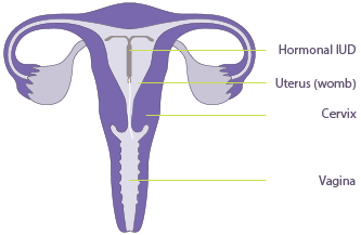
An Intrauterine Device (IUD) is often prescribed to women for contraception purposes or to control heavy periods. Commonly known as the Mirena (or Kyleena), this hormonal device is inserted into the uterus for a long-term effect.
At Adelaide Women’s Imaging (AWI), we offer women the option to guide the Mirena into the uterus using Ultrasound.
Ultrasound guided Mirena insertion is particularly useful for women who have never given birth, have had a previous caesarian section, and those with a retroverted uterus (a uterus that is tipped backwards).
Ultrasound guidance allows optimal placement of the Mirena within the uterus cavity and reduces the risk of perforation. After the insertion of the Mirena, an ultrasound image is taken to confirm the placement of the device. Using ultrasound guidance for Mirena insertion may also be better tolerated by women by reducing the need to use a more invasive technique to gain access to the cervix.
Ultrasound guidance can also be used to remove an IUD if necessary.
Before Your Ultrasound Guided Mirena Insertion
Please ensure that your doctor has done vaginal swabs to exclude infection before the procedure. Please try and bring these results with you.
Empty your bladder 1 hour prior to the procedure, then drink 2 glasses (600ml) of water and hold it. Do not empty your bladder again. Your Doctor should give you a script for the Mirena/Kyleena which you must get filled and bring to the appointment. Adelaide Women’s Imaging will not provide the device.
Procedure
When you visit our clinic for a Mirena/Kyleena removal or insertion, you will be greeted by one of our friendly reception staff. A sonographer will then collect you from the waiting room and take you to the ultrasound room. The sonographer will perform an ultrasound of the abdomen (transabdominal ultrasound) to specifically examine the uterus and ovaries to determine the shape and size of your uterus.
We will then ask you to partially empty your bladder for a urinary pregnancy test. When the scan has been completed, the doctor will insert a speculum; a medical instrument to separate the walls of the vagina, similar to when you have a PAP smear.
 The initial set of ultrasound images taken by the sonographer will be reviewed by an AWI obstetrician and gynaecologist. The doctor will perform the procedure, and feedback will be given to you at the time of your appointment.
After Your Scan
Other than mild discomfort, complications are rare. Some women may experience mild cramping or bleeding for a few days. Pelvic infection following the procedure is extremely rare, but you should report any symptoms of pain, tenderness, fever or excessive vaginal discharge to your doctor should this occur.
Duration
The procedure will take approximately 45 minutes.

